SKT-120-96S SKT-120-480S
MOLECULAR SIGNATURE® DNA Damage (8-OHdG) ELISA Kit
Stressmarq Biosciences
Image
Image
Image
Image
Image
Image
Image
DETAILS
- Item: 8-hydroxy-2-deoxy Guanosine : BSA Coated Plate | 8-hydroxy-2-deoxy Guanosine Standard | 8-hydroxy-2-deoxy Guanosine HRP Conjugated Monoclonal Antibody | Sample and Standard Diluent | 8-hydroxy-2-deoxy Guanosine Antibody Diluent | Wash Buffer Concentrate | TMB Substrate | Stop Solution | Plate Cover
- Target: DNA Damage (8-OHdG)
- Utility: ELISA Kit for 8-OHdG detection in samples.
- Category: Assay Kits
- Platform: Microplate
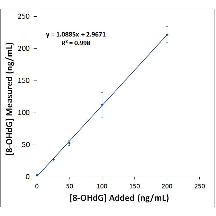
- Precision: Intra-Assay Precision: Three samples of known concentration were assayed thirty times on one plate; the intra-assay coefficient of variation of the DNA Damage ELISA has been determined to be <5%. Inter-Assay Precision: Three samples of known concentration were assayed thirty times in three individual assays; the inter-assay coefficient of variation of the DNA Damage ELISA has been determined to be <5%.
- Assay Type: Competitive ELISA (Enzyme-linked Immunosorbent Assay)
- References: 1. Maxey K.M., Maddipati K.R., Birkmeier J. (1992) J Clin Immunoassay 15: 116-120. 2. Pradelles P., Grassi J., Maclouf J. (1990) Methods Enzymol. 187: 24-34. 3. Maclouf J., Grassi J., Pradelles P. (1987) Dev Immunoassay Tech Meas eicosanoids. 4. Lin H., et al. (2004) Biochem J. 380: 541-548. 5. Bogdanov M.B., et al. (1999) Free Radic Biol Med. 27(5/6): 647-666. 6. Lee J., et al. (2005) Hypertension 45: 986-990. 7. Leinonen, J., et al. (1997) FEBSLett. 417: 150-152. 8. Endo K., et al. (2006) J. Atheroscler. Thromb. 13:68-75. 9. Kuo H., et al. (2007) Mutat Res. 631:62-68. 10. Shen J., et al. (2007) Cancer 109: 574-580. 11. Beckman K.B., Ames B.N. (1997) J Biol Chem 272: 19633-19636. 12. Epe B., et al. (1996) Nucleic Acids Res 24: 4105-4110. 13. Spencer J.P.E., et al. (1995) FEBS Lett 374: 233-236. 14. Floyd R.A. (1990) FASEB J 4: 2587-2597.
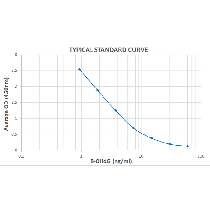
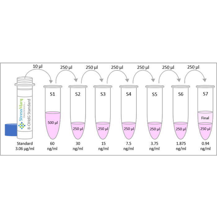
- Assay Range: 0.94 - 60 ng/mL
- Description: Colorimetric detection of 8-hydroxy-2-deoxy Guanosine
- Sensitivity: 0.59 ng/mL
- Specificity: Cross-reactivity: 8-Hydroxy-2-deoxy Guanosine (8-OHdG): 100%. 8-Hydroxy Guanosine (8-OHG): 23%. 8-Hydroxy Guanine (8-oxoG): 23%. Guanosine: <0.01%.
- Component No: SKC-120A | SKC-120C | SKC-120F | SKC-0001 | SKC-0002 | SKC-0003 | SKC-0004 | SKC-0005 | SKC-0009
- Field of Use: Not for use in humans. Not for use in diagnostics or therapeutics. For in vitro research use only.
- Net Weight G: 500
- Product Type: ELISA Kits
- Sample Types: Urine | Cell Lysates | Plasma | Sample matrices
- Stock Product: Y
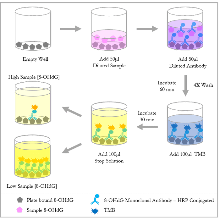
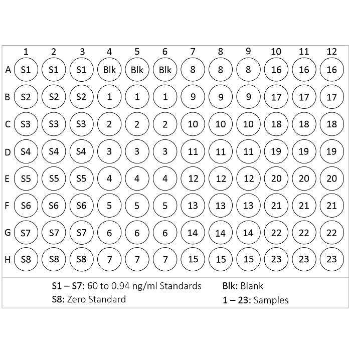
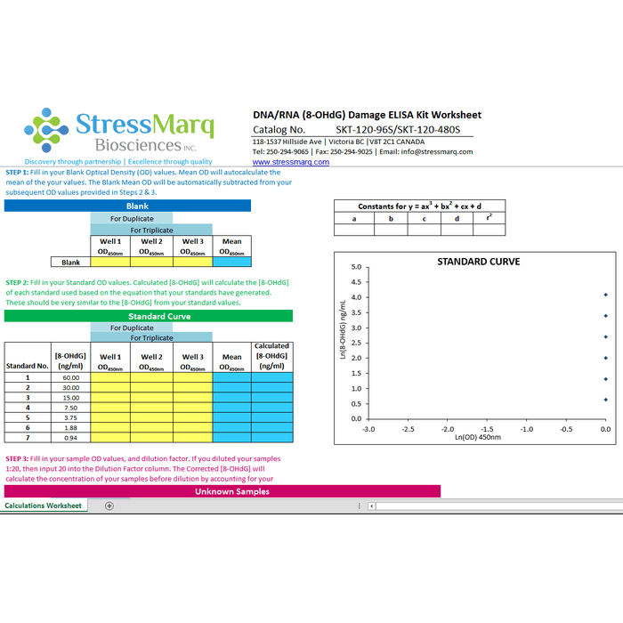
- Assay Overview: 1. Prepare standard and samples in the Sample and Standard Diluent. 2. Add 50 µL of prepared standards and samples in triplicate to appropriate wells. 3. Add 50 µL of the diluted antibody preparation to the appropriate wells. 4. Cover plate with Plate Cover and incubate at room temperature (20-25°C) for 1 hour. 5. Wash plate 4 times with 1X Wash Buffer. 6. Add 100 µL of TMB Substrate to each well. 7. Cover plate and develop the plate in the dark at room temperature for 30 minutes. 8. Add 100 µL of Stop Solution to each well. 9. Measure absorbance on a plate reader at 450 nm. 10. Plot the standard curve and calculate sample concentrations.
- Quantity Size: 1 Plate | 1 vial/ 100uL | 1 vial/75uL | 1 vial/50mL | 1 vial/13mL | 1 vial/50mL | 1 vial/13mL | 1 vial/13mL | 2 covers
- Research Areas: Cancer | Oxidative Stress | Cell Signaling | Post-translational Modifications | Oxidation | Cell Signaling | Epigenetics and Nuclear Signaling | DNA/RNA | DNA Damage and Repair
- Gross Weight Kg: 2
- Incubation Time: 1 hour
- Detection Method: Colorimetric Assay
- Alternative Names: 8-OH-dG ELISA Kit, 8OHG ELISA Kit, 80G ELISA Kit, 8 hydroxyguanine ELISA Kit, 8-OHdG ELISA Kit, DNA Damage ELISA Kit
- Cite This Product: DNA Damage (8-OHdG) ELISA Kit (StressMarq Biosciences, Canada, Cat # SKT-120-96S)
- Country of Origin: Canada
- Number of Samples: 39 samples in duplicate
- Storage Temperature: 4ºC and -20ºC
- Shipping Temperature: Blue Ice
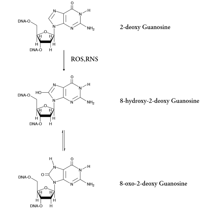
- Scientific Background: 8-hydroxy-2-deoxy Guanosine (8-OH-dG) is produced by the oxidative damage of DNA by reactive oxygen and nitrogen species and serves as an established marker of oxidative stress (1-4). Hydroxylation of guanosine occurs in response to both normal metabolic processes and a variety of environmental factors (i.e., anything that increases reactive oxygen and nitrogen species). Increased levels of 8-OH-dG are associated with the aging process as well as with a number of pathological conditions including cancer, diabetes, and hypertension(5-9). In complex samples such as plasma, cell lysates, and tissues, 8-OH-dG can exist as either the free nucleoside or incorporated in DNA. Once the blood enters the kidney, free 8-OH-dG is readily filtered into the urine, while larger DNA fragments remain in the bloodstream. Because of the complexity of plasma samples, urine is a more suitable matrix for the measurement of free 8-OH-dG than plasma. Urinary levels of 8-OH-dG range between 2.7-13 ng/mg creatine, while plasma levels of free 8-OH-dG have been reported to be between 4-21 pg/ml as determined by LC-MS (10-11).
- Species Reactivity Medium: Species Independent
- Species Reactivity Full Name: Species Independent
DESCRIPTION
Colorimetric detection of 8-hydroxy-2-deoxy Guanosine