SKT-105-96 SKT-105-480
StressXpress® HSP70 ELISA Kit
Stressmarq Biosciences
Image
DETAILS
- Item: Anti-Hsp70 Immunoassay Plate | 5X Hsp70 Extraction Reagent | Recombinant Hsp70 Standard | Standard and Sample Diluent | 10X Wash Buffer Concentrate | Anti-Hsp70 Biotinylated Antibody Concentrate | Anti-Hsp70 Biotinylated Antibody Diluent | Streptavidin: HRP Concentrate | Streptavidin: HRP Diluent | TMB Substrate | Stop Solution | Pre-treatment Buffer
- Target: HSP70
- Utility: ELISA kit used to quantitate HSP70 concentration in samples.
- Category: Assay Kits
- Platform: Microplate
- Assay Type: Sandwich ELISA (Enzyme-linked Immunosorbent Assay)
- References: 1. Zho J. (1998) Cell. 94: 471-480. 2. Boorstein W.R., Ziegelhoffer T. & Craig E.A. (1993) J. Mol. Evol. 38(1): 1-17. 3. Rothman J. (1989) Cell. 59: 591 -601. 4. DeLuca-Flaherty et al. (1990) Cell. 62: 875-887. 5. Bork P., Sander C. & Valencia A. (1992) Proc. Natl Acad. Sci. USA. 89: 7290-7294. 6. Fink A.L. (1999) Physiol. Rev. 79: 425-449. 7. Smith D.F., et al. (1993) Mol. Cell. Biol. 13(2): 869-876. 8. Prapapanich V., et al., (1996) Mol. Cell. Biol. 16(11): 6200-6207. 9. Fernandez-Funez et al., (2000) Nature. 408(6808): 101-106.
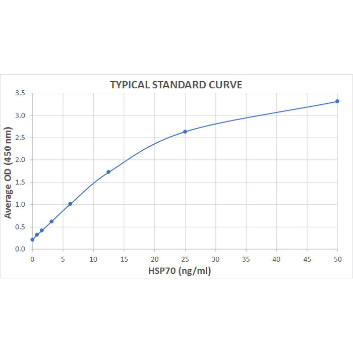
- Assay Range: 0.781 - 50 ng/ml
- Description: Colorimetric detection of HSP70
- Sensitivity: 0.18 ng/ml
- Component No: SKC-105A | SKC-105B | SKC-105C | SKC-105D | SKC-105E | SKC-105F | SKC-105G | SKC-105H | SKC-105I | SKC-105J | SKC-105K | SKC-105L
- Field of Use: Not for use in humans. Not for use in diagnostics or therapeutics. For in vitro research use only.
- Net Weight G: 500
- Product Type: ELISA Kits
- Sample Types: Cell Lysates | Tissue | Urine | Saliva
- Stock Product: Y
- Assay Overview: 1. Prepare Standard and samples in Standard and Sample Diluent. 2. Add 100 µL of Standard to appropriate wells. 3. Add 50 µL of Pre-Treatment Buffer to all sample wells. 4. Add 50 µL of sample to appropriate wells. 5. Cover plate with Plate Sealer and incubate at 37°C for 2 hours. 6. Wash plate four times with 1X Wash Buffer. 7. Add 100 µL of Detection Antibody Working Solution to each well. 8. Cover plate with Plate Sealer and incubate at 37°C for 2 hours. 9. Wash plate four times with 1X Wash Buffer as described in step 6. 10. Add 100 µL of Streptavidin-HRP Working Solution to each well. 11. Cover plate with Plate Sealer and incubate at room temperature for 30 minutes. 12. Wash plate four times with 1X Wash Buffer as described in step 6. 13. Add 100 µL of TMB Substrate to each well. 14. Develop the plate in the dark at room temperature for 30 minutes. 15. Stop reaction by adding 100 µL of Stop Solution to each well. 16. Measure absorbance on a plate reader at 450 nm.
- Quantity Size: 1 Plate | 1 vial/10 ml | 2 vials | 1 vial/ 50 ml | 1 vial/100 ml | 1 vial/150 µl | 1 vial/ 13 ml | 1 vial/150 µl | 1 vial/ 13 ml | 1 vial/ 13 ml | 1 vial/ 13 ml | 1 vial/ 13 ml
- Research Areas: Cancer | Heat Shock | Cell Signaling | Protein Trafficking | Chaperone Proteins | Cancer | Tumor Biomarkers
- Gross Weight Kg: 2
- Incubation Time: 30 minutes
- Detection Method: Colorimetric Assay
- Alternative Names: HSP70 1 ELISA Kit, HSP70 2 ELISA Kit, HSP70.1 ELISA Kit, HSP72 ELISA Kit, HSPA1 ELISA Kit, HSPA1A ELISA Kit, HSPA1B ELISA Kit
- Cite This Product: HSP70 ELISA Kit (StressMarq Biosciences, Canada, Cat # SKT-105-96)
- Country of Origin: Canada
- Number of Samples: 40 samples in duplicate
- Storage Temperature: 4ºC and -20ºC
- Shipping Temperature: Blue Ice
- Scientific Background: HSP70 genes encode abundant heat-inducible 70-kDa HSPs (HSP70s). In most eukaryotes HSP70 genes exist as part of a multigene family. They are found in most cellular compartments of eukaryotes including nuclei, mitochondria, chloroplasts, the endoplasmic reticulum and the cytosol, as well as in bacteria. The genes show a high degree of conservation, having at least 5O% identity (2). The N-terminal two thirds of HSP70s are more conserved than the C-terminal third. HSP70 binds ATP with high affinity and possesses a weak ATPase activity which can be stimulated by binding to unfolded proteins and synthetic peptides (3). When HSC70 (constitutively expressed) present in mammalian cells was truncated, ATP binding activity was found to reside in an N-terminal fragment of 44kDa which lacked peptide binding capacity. Polypeptide binding ability therefore resided within the C-terminal half (4). The structure of this ATP binding domain displays multiple features of nucleotide binding proteins (5). All HSP70s, regardless of location, bind proteins, particularly unfolded ones. The molecular chaperones of the HSP70 family recognize and bind to nascent polypeptide chains as well as partially folded intermediates of proteins preventing their aggregation and misfolding. The binding of ATP triggers a critical conformational change leading to the release of the bound substrate protein (6). The universal ability of HSP70s to undergo cycles of binding to and release from hydrophobic stretches of partially unfolded proteins determines their role in a great variety of vital intracellular functions such as protein synthesis, protein folding and oligomerization and protein transport. Looking for more information on HSP70? Visit our new HSP70 Scientific Resource Guide at http://www.HSP70.com.
- Species Reactivity Medium: Human | Goat | Dog | Rat
- Species Reactivity Full Name: Human | Goat | Dog | Rat
DESCRIPTION
Colorimetric detection of HSP70